68 Knee Anatomy With Nerves
The common peroneal nerve diverges laterally running just behind the tendon of biceps femoris. The common peroneal nerve diverges laterally running just behind the tendon of biceps femoris.

Geniculate Artery Embolization In Patients With Recurrent Hemarthrosis After Knee Arthroplasty A Retrospective Study The Journal Of Arthroplasty
The important parts of the knee include.
68 knee anatomy with nerves. Free full text J Ultrasound. The knee is the meeting place of two important bones in the leg the femur the thighbone and the tibia the shinbone. Now i have a pinched nerve in my knee.
US and MR Imaging with Anatomic Correlation1 The anatomy of the nerves of the foot and ankle is complex and familiarity with the normal anatomy and course of these nerves as well as common anatomic variants is essential for correct identifi-cation at imaging. Normal Anatomy and Compres-sion Areas of Nerves of the Foot and Ankle. Various nerves and blood vessels supply the muscles and bones of the knee.
Nerves in knees anatomy. The large sciatic nerve splits just above the knee to form the tibial nerve and the common peroneal nerve. The anatomy of the knee knee bones knee muscles knee arteries knee veins and nerves looking into the anatomy of the knee.
BackgroundDespite their emerging therapeutic relevance there are many discrepancies in anatomical description and terminology of the articular nerves supplying the human knee capsule. In this page we also recommend where to buy best selling health care products at a lower price. Nerves In The Knee Anatomy.
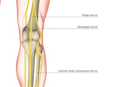
Medial sural cutaneous nerve. Nervesthe two plexi that contribute to the nervous innervation of the lower limb are the lumbar plexus and sacral plexus. Agony from more profound damage called alluded torment can be passed along the nerve to be felt superficially.
Hailey Daniswicz flexes muscles in her thigh in Chicago April 13 2011 as electrodes attached to her leg instruct a computer avatar to flex its knee and ankle - parts of. The posterior knee joint was innervated by two or three nerves most commonly via. Learn vocabulary terms and more with flashcards games and other study tools.
The Tibial nerve from the anterior divisions of L4-S3. Search from Knee Nerves stock photos pictures and royalty-free images from iStock. Anatomy of the knee knee bones knee muscles knee arteries knee veins and nerves looking into the anatomy of the knee.
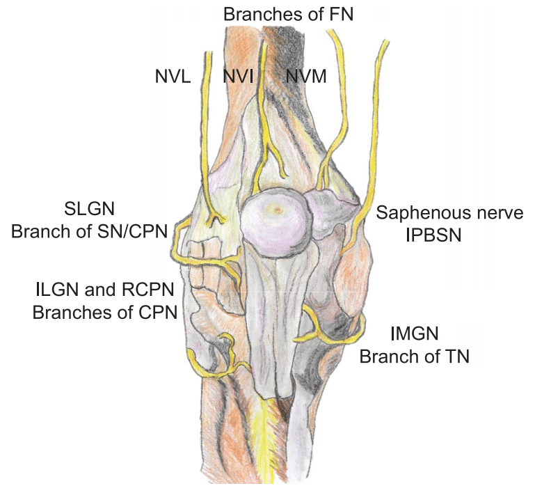
Branches of major nerves5 Knee sensory innervation The sensory innervation of the knee is basically divided into two groups of nerves the posterior group and the anterior group6 The posterior group originates from the sciatic nerve mainly by its posterior tibial branch upper medial geniculate nerves lower and. Knee Anatomy Osteology Arthrology Ligaments Muscles And Nerves 01 Knee Anatomy. The knee is designed to fulfill a number of.
The most important nerves around the knee are the tibial nerve and the common peroneal nerve in the back of the knee. Find high-quality stock photos that you wont find anywhere else. Knee anatomy with nerves.
The store that we recommend also provides. Surgical and Radiologic Anatomy. The knee is one of the largest and most complex joints in the body.
Daniswicz is part of a clinical trial sponsored by the US. The most important nerves around the knee are the tibial nerve and the common peroneal nerve in the back of the knee. Tendons connect the knee bones to the leg muscles that move the knee joint.
The patella or kneecap as it is commonly called is made of bone and sits in front of the knee. The knee is designed to fulfill a number of functions. The most important nerves around the knee are the tibial nerve and the common peroneal nerve in the back of the knee.
Above the knee the sciatic nerve divides into two major nerves the tibial nerve and the common peroneal nerve. This nerve branches off the sciatic nerve in the popliteal fossa and runs along the biceps femoris and leaves the fossa to run around the head of the fibula and down the leg to the ankle. Figure 2 From Treatment Of Peroneal Nerve Injuries In The The lumbar plexus l1 5 gives rise to the femoral and obturator nerves that innervate the hip flexors and adductors and the knee extensors.
The large sciatic nerve splits just above the knee to form the tibial nerve and the common peroneal nerve. Start studying Knee Anatomy. If you are looking for Nerves In The Knee Anatomy youve come to the right place.
The knee joint is a synovial joint. The anterior knee joint was innervated by 10 nerves and further subdivided into two parts anteromedial and anterolateral or four quadrants superomedial inferomedial superolateral and inferolateral based on innervation patterns. The most important nerves around the knee are the tibial nerve and the common peroneal nerve in the back of the knee.
Beneath the fascia lata. Army that is using electromyogr. Lets look at a normal knee joint to understand how the parts anatomy work together function and how knee.
With the knee bent a doctor can pull anterior drawer test and push posterior drawer test the lower leg while holding the foot stable to check the stability of the acl and pcl. In this article we will review the normal gross and microscopic anatomy of the nerves in the knee region the US technique used for their examination and their normal US appearance. And it supplies the posterior thigh posterior leg and plantar muscles and.
Anatomy of the knee bones muscles arteries veins nerves bone and ligaments. Support the body in an upright position without the need for muscles to work. Each type of nerve is the relay for pain signals that originate in a different area of your knee.
Full list of the names of bones muscles veins arteries and veins found in the knee. Knee torment along these lines can emerge from the knee itself or be alluded from states of the hip lower leg or lower back. These two nerves travel to the lower leg and foot supplying sensation and muscle control.
All of us review 9 related goods including videos deals discount coupon pictures and more. This cadaveric study aimed to determine their origin trajectory relationship and landmarks for therapeutic purposeMethodsWe. The nerves that give sensation to the knee return from the lower and furthermore give hip leg and more moderate leg sensation.
The sciatic nerve travels down the thigh to the area of the popliteal fossa and at this point it divides into the tibial and common peroneal nerves. The superior gluteal nerve L4-S1 goes to the gluteus medius gluteus minimus and tensor fasciae latae The inferior gluteal nerve L5-S2 goes to the gluteus maximus The sciatic nerve which is the largest nerve in the body. The anatomy of the knee knee bones knee muscles knee arteries knee veins and nerves looking into the anatomy of the knee.
Is formed of two parts.

The Superficial Anatomy Of The Medial Knee Modified From Kim By Author Download Scientific Diagram

Peripheral Nerve Block Flos Block For Intraoperative Anesthesia In Total Knee Arthroplasty An Observational Study Khanna S Gogoi B Jaishree Sv Prasad G V Indian J Health Sci Biomed Res

The Superficial Anatomy Of The Medial Knee Modified From Kim By Author Download Scientific Diagram

Laminated Anatomy And Injuries Of The Knee Poster Knee Joint Anatomical Chart 18 X 27 Amazon Com Industrial Scientific

Anatomical Landmark For Genicular Nerves The Targets Included The Sm Download Scientific Diagram
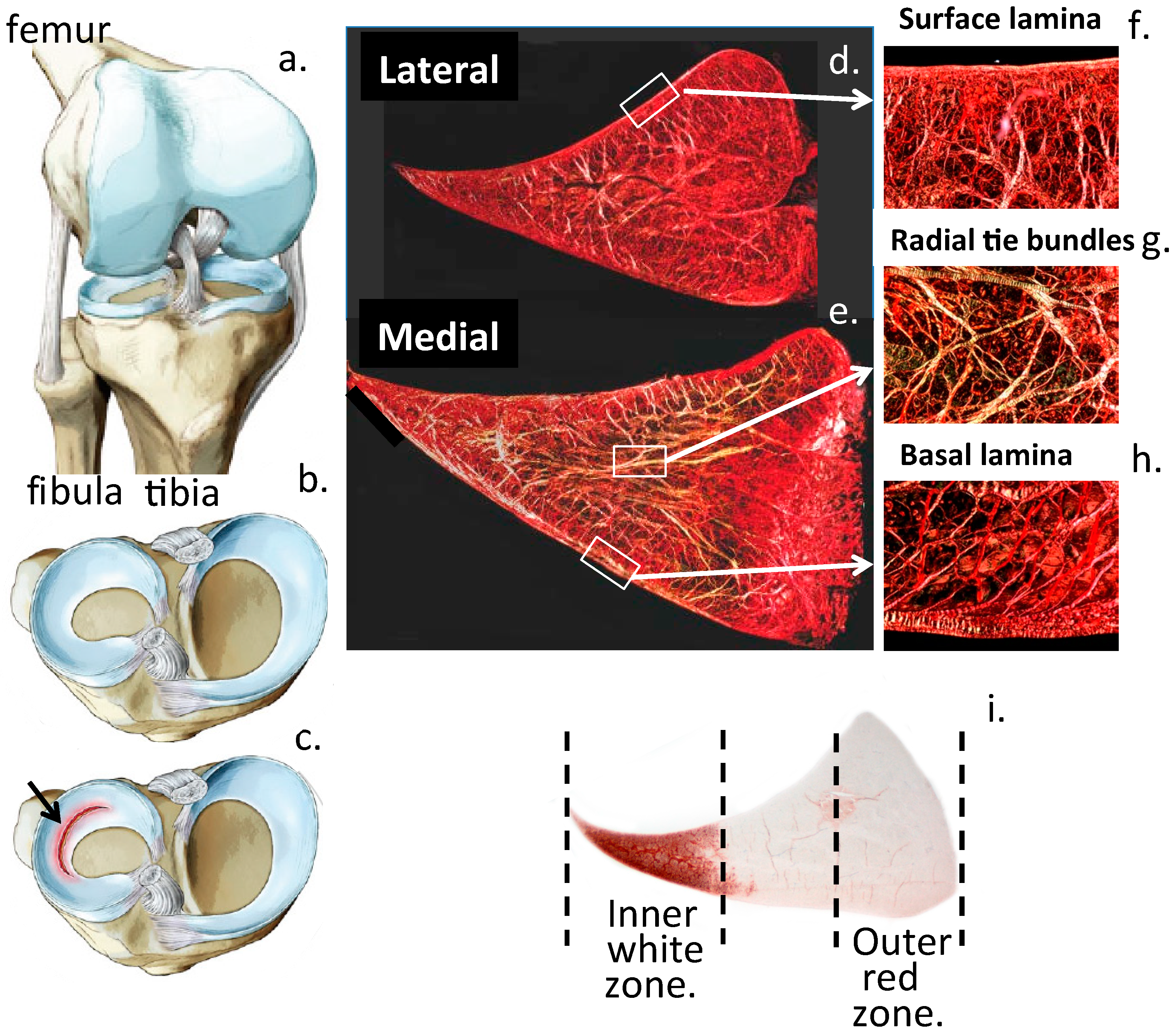
Cells Free Full Text The Importance Of The Knee Joint Meniscal Fibrocartilages As Stabilizing Weight Bearing Structures Providing Global Protection To Human Knee Joint Tissues Html

Clinical Anatomy Of Lower Extremity Basicmedical Key
Anatomy Of Back View Of Knee Stock Photos Page 1 Masterfile

Obturator Nerve Block Landmarks And Nerve Stimulator Technique Nysora

Pin By Klausbahl On Genu Valgum Treatment Medical Anatomy Muscle Anatomy Human Anatomy And Physiology
Motor Sparing Blocks For The Knee Wfsa Resources

Anatomy Musculoskeletal Key
Knee Joint Intra Articular Injection Anesthesia Key

Anatomy Of The Knee Bones Muscles Arteries Veins Nerves Anatomy Of The Knee Joints Anatomy Human Anatomy And Physiology

The Anatomy And Pattern Of Pain Of The Saphenous Nerve At The Knee Download Scientific Diagram

Regional Analgesia For Knee Surgeries Thinking Beyond Borders Intechopen

Anatomy Of The Knee Bones Muscles Arteries Veins Nerves Anatomy Of The Knee Human Anatomy And Physiology Medical Anatomy

Surface Landmarks Of The Ips Download Scientific Diagram

Anatomy Of Human Knee Joint Poster By Stocktrekimages In 2021 Human Knee Knee Muscles Anatomy Shoulder Anatomy