Left Knee Anatomy Posterior View
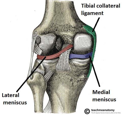
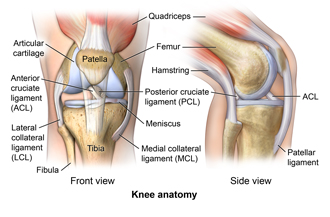
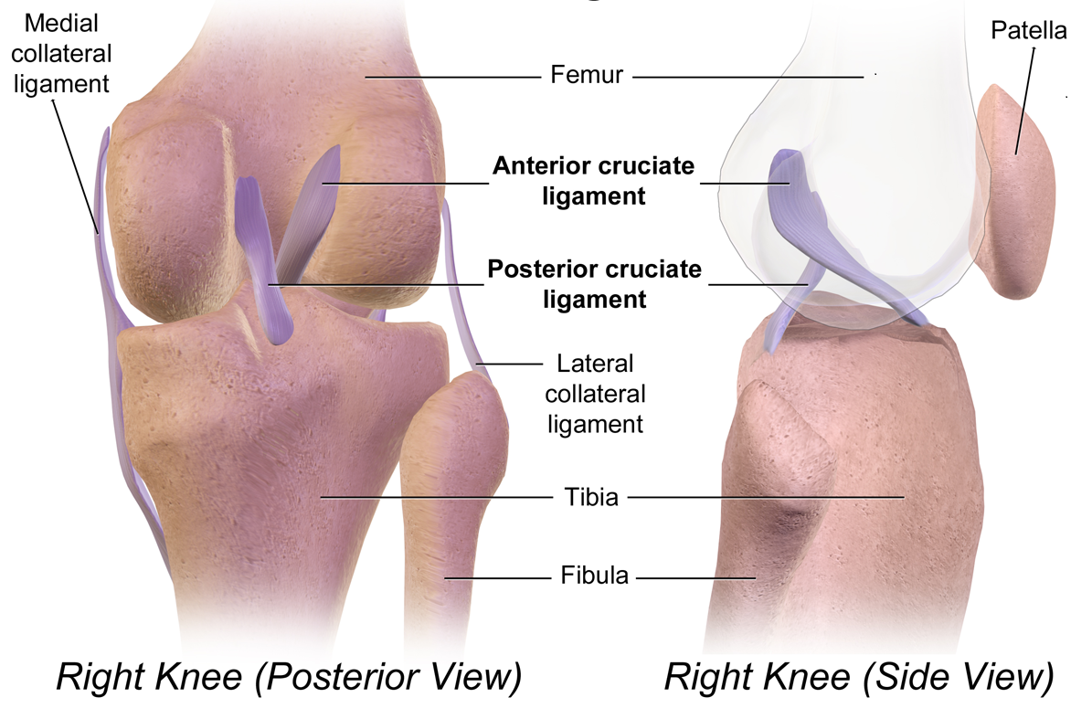
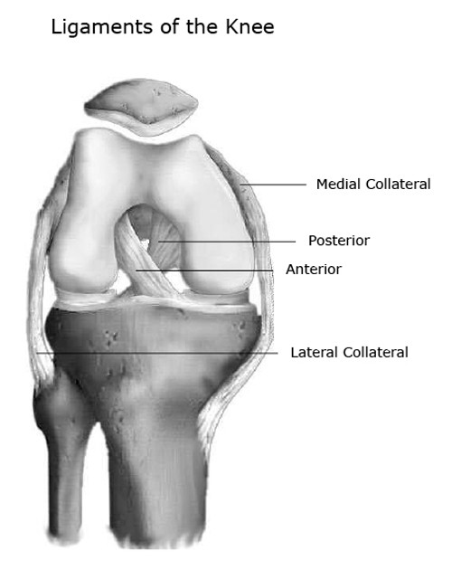
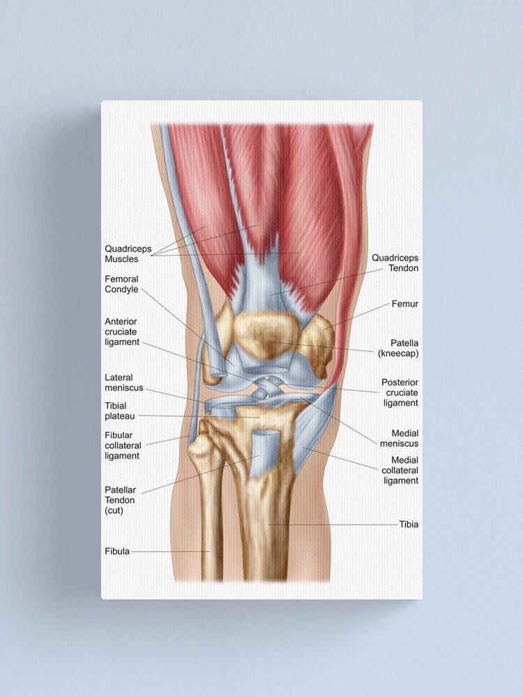
Increased posterior translation on the posterior drawer test indicates a combined posterior cruciate ligament tear with the pcl injury. One ligament is on each side of the knee jointthe medial collateral ligament on the inner side and the lateral collateral ligament on the outer side.
 Posterior View Of Human Left Knee Joint 5 Download Scientific Diagram
Spin echo t1 or proton density with fat saturation sequences.

Left knee anatomy posterior view. Gradual onset or chronic knee pain develops over time and is often caused by overuse. Pcl posterior cruciate ligament strain or tear. An alternative to the supine position is the standing ap image.
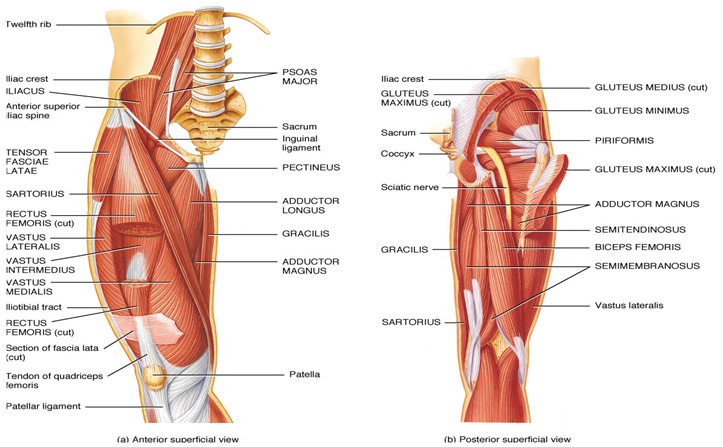
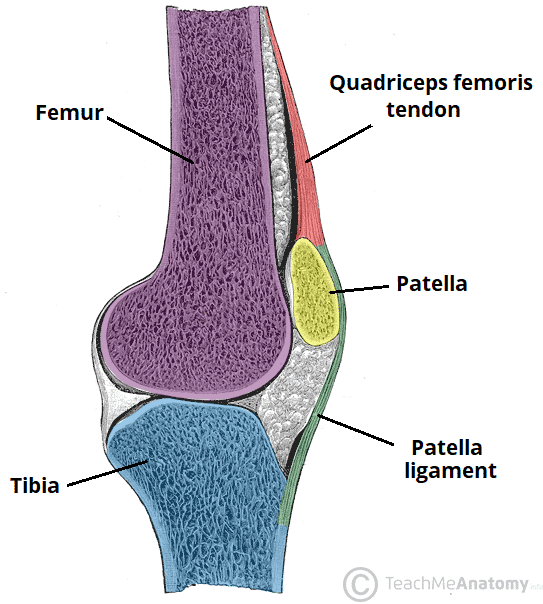
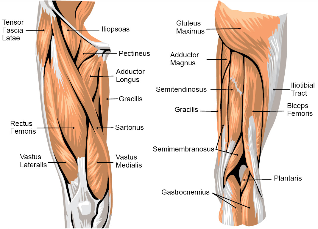
Flat muscle enabling the thigh to extend on the pelvis the knee to flex and the thigh and the leg to rotate inwardly toward the median axis. The knee is the meeting point of the femur thigh bone in the upper leg and the tibia shinbone in the. Anatomy of the knee on a coronal slice mri.
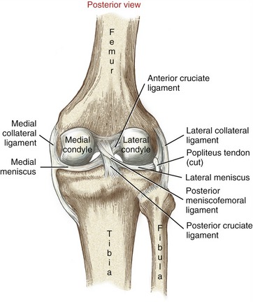
These injuries are less common than acl tears and physical therapy rather than surgery. You will also find anterior cruciate ligament lateral epicondyle posterior meniscofemoral ligament. Pcl tears can cause pain swelling and knee instability.
Posterior knee pain is a common patient complaint. Posterior view of knee joint. The posterior cruciate ligament prevents the femur from.
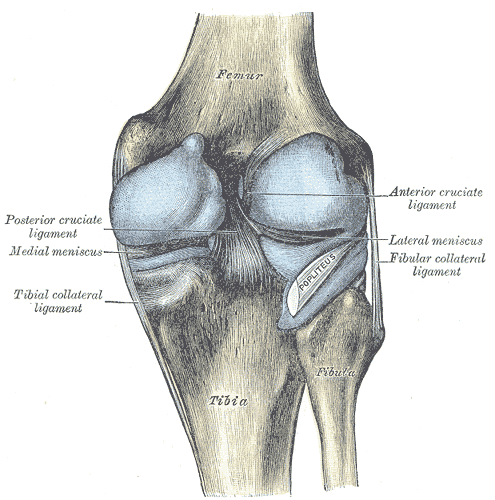
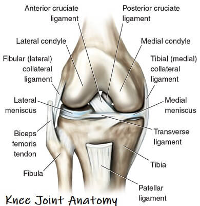
Medical images from an mri allow medical professionals to distinguish body tissues including the meniscus shock absorbers in the knee cartilage tendons and ligaments. Semitendinosus long muscle enabling the thigh to extend on the pelvis the knee to flex and the thigh and the leg to rotate inwardly toward the median axis. These are called the cruciate ligaments and consist of the anterior cruciate ligament and the posterior cruciate ligament.
The knee is a complex joint that flexes extends and twists slightly from side to side. In supine position the x rays pass through the knee from anterior to posterior ap image. Posterior view of knee joint.
Magnetic resonance imaging mri is a radiologic procedure that uses a magnetic field and radio waves to develop detailed image cross sections of the body including the knee 1. In this image you will find intercondylar notch medial epicondyle medial meniscus tibial collateral ligament posterior cruciate ligament the popliteal surface of the tibia in it. On contrast the user can choose the type of mri sequence.
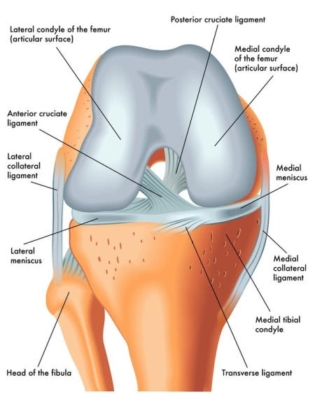
Meniscus lateral and medial cruciate ligaments vastus lateralis intermedius medialis tibial and fibular collateral ligaments. The front to back or anterior posterior knee image can be made in both supine and standing positions fig.

 Posterior View Of A Left Leg Mapping The Location Of The Different Muscles That Make It Up Human Body Anatomy Human Muscle Anatomy Muscle Anatomy
 Structure And Function Of The Knee Musculoskeletal Key
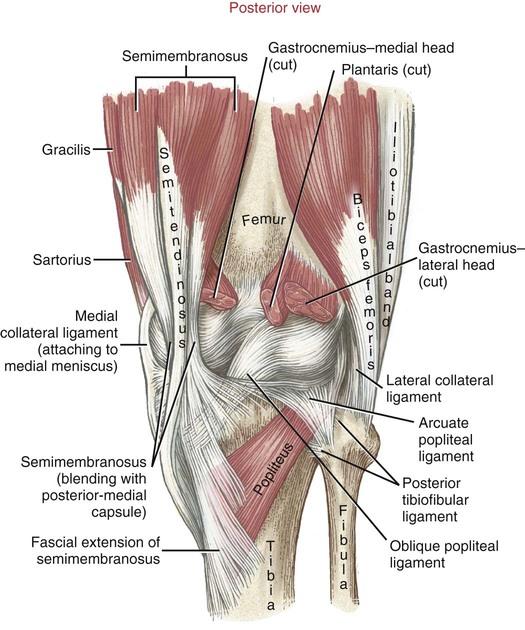
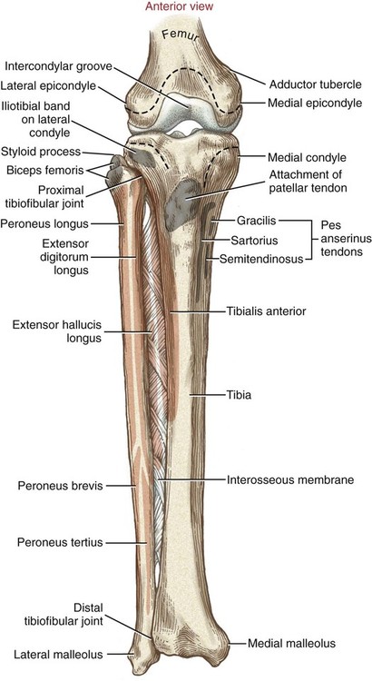
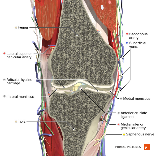
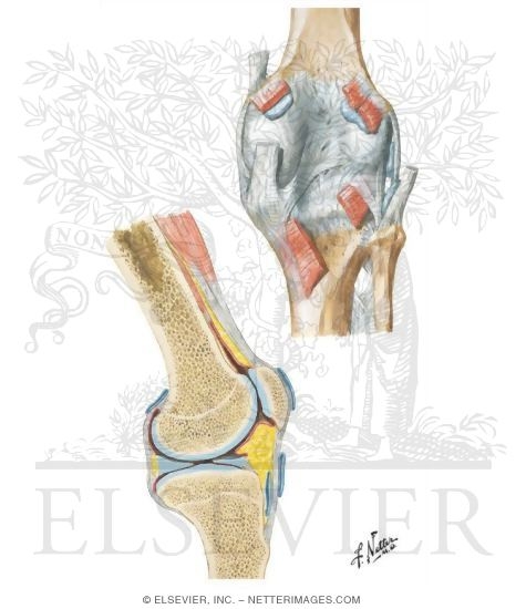
 Knee Leg Atlas Of Anatomy
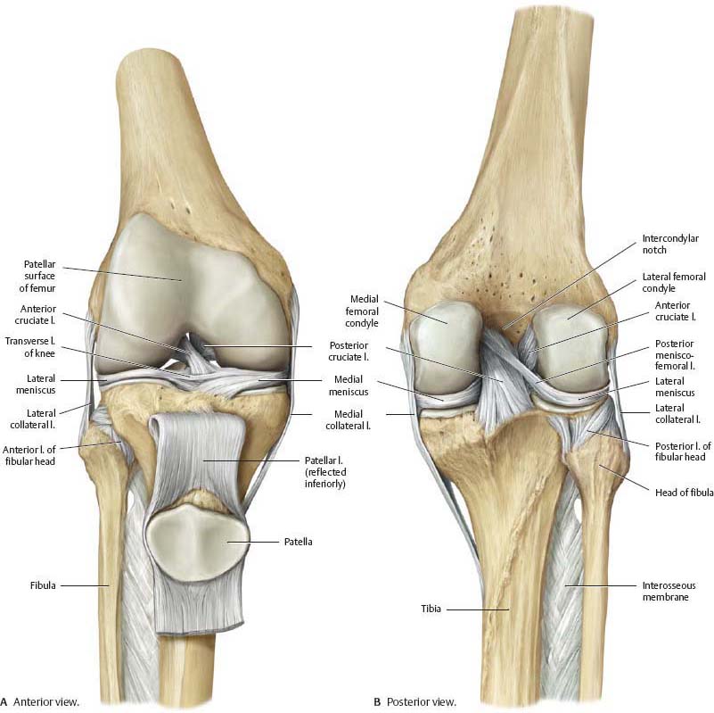
 Knee Anatomy Anterior View And Posterior View
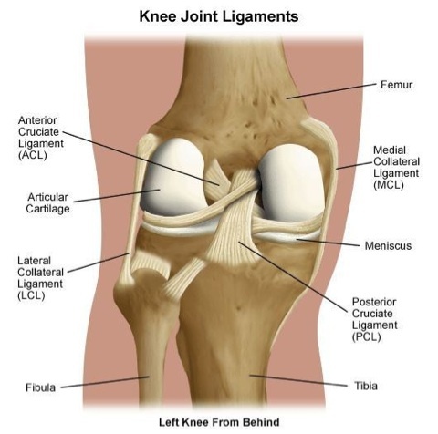 Types Of Knee Ligaments Stanford Health Care
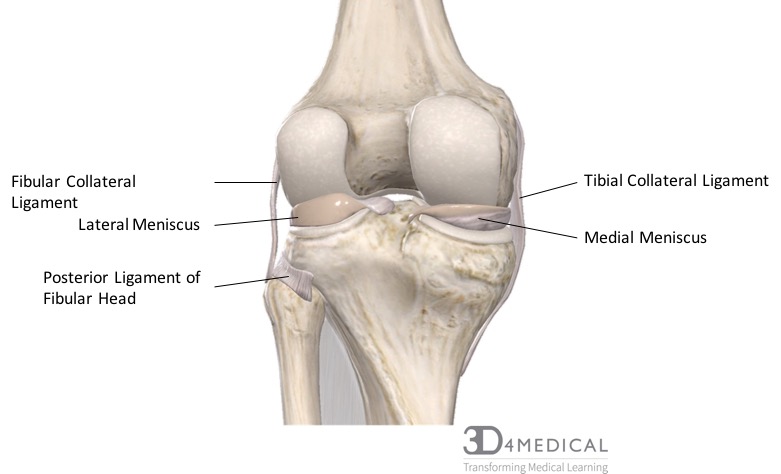
 Posterior View Of Knee Joint

 Knee Leg Atlas Of Anatomy
 Posterior View Of The Right Knee Anatomy Of The Posterior Of The Right Knee In Extension Canstock
 Anatomy Of The Left Knee Medical Illustration Medivisuals
/188058334-crop-56aae7425f9b58b7d0091480.jpg) What Is Causing Your Knee Pain
What Is Causing Your Knee Pain
 Joints Ligaments And Connective Tissues Advanced Anatomy 2nd Ed
 Pcl Injury Knee Sports Orthobullets
 Anatomy Of The Knee Bones Muscles Arteries Veins Nerves Anatomy Of The Knee Bones And Muscles Knee Bones

 Adult Knee Radiographic Evaluation Recon Orthobullets
 Posterior Knee Pain Physiopedia
 Knee Clinical Gate
 The Knee Joint Articulations Movements Injuries Teachmeanatomy
 Knee Wikipedia
 Posterior View Of The Knee With Posterior Capsules Shown In Blue Download Scientific Diagram
 Knee Anatomy Exhibits
Https Encrypted Tbn0 Gstatic Com Images Q Tbn And9gcsswc 7weyyuu6rmyszxnmvyzxzyc0c1deffntz18oeb1cninyc Usqp Cau

 Knee Injury Treatment Hurt911
![]() Leg And Knee Anatomy Bones Muscles Soft Tissues Kenhub
Leg And Knee Anatomy Bones Muscles Soft Tissues Kenhub
What Is The Proper Name For The Back Of The Knee Quora
 Muscle Charts Massagelongbeachca Com
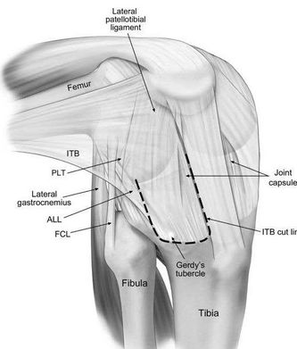
![]() Anterolateral Ligament Newly Described Ligament Kenhub
Anterolateral Ligament Newly Described Ligament Kenhub
 Leg Definition Bones Muscles Facts Britannica
 Normal Left Knee Anatomy Meniscus Medical Art Works
 Left Knee Joint In Flexion And Torn Acl
 Illustration Of Left Knee Medial View Showing Bones Tendons Muscles Meniscus And Ligaments With The Medial Patellar Retinaculum Retracted Orthopaedic Surgical Anatomy Teaching Collection Usc Libraries Digital Collections
 Pin On Just Me
 Knee Wikipedia
![]() N4welpbzucz0xm
N4welpbzucz0xm
 Knee Joint Anatomy Motion Knee Pain Explained
 The Knee Joint Articulations Movements Injuries Teachmeanatomy
 Posterior View Of A Left Foot And Ankle In Neutral Plantar Flexion And Download Scientific Diagram
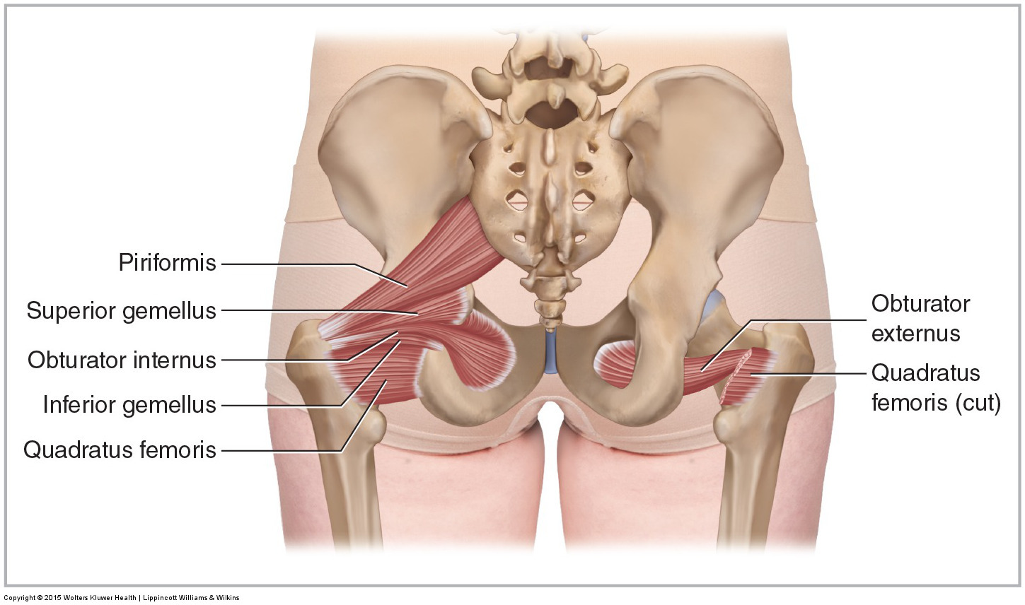 Muscles Of The Pelvis
 Is The Knee In Danger In A Figure 4 Position Sara Doyle Yoga And Anatomy
 Muscles Of The Knee Anatomy Pictures And Information

 Knee Injuries
 Structure And Function Of The Knee Musculoskeletal Key
 Keeping On Track With Knees Expanding Light
Https Encrypted Tbn0 Gstatic Com Images Q Tbn And9gct6olqr 6gaygjdc2mmixncssaguqyuoqzfg5otz Icphxxhtuv Usqp Cau
 Knee Joint Anatomy Bones Ligaments Muscles Tendons Function
 Normal Posterior Left Knee Anatomy Medical Art Works

 Types Of Knee Pain Anterior Posterior Medial Lateral
 Knee Ultrasound Radiology Key
Common Knee Injuries Orthoinfo Aaos
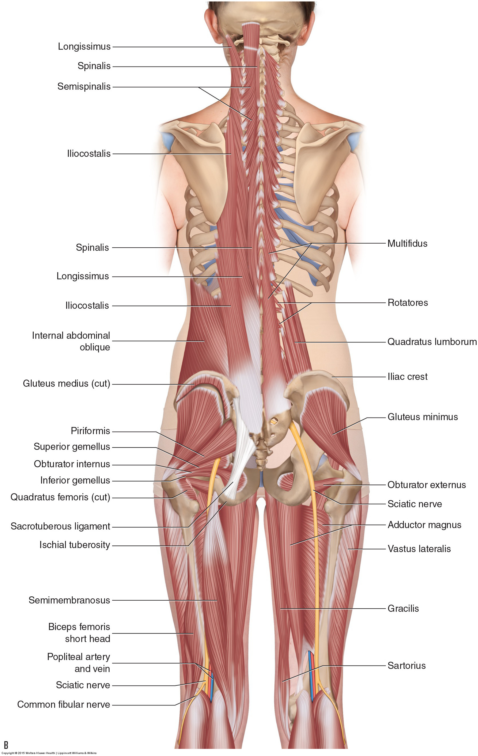
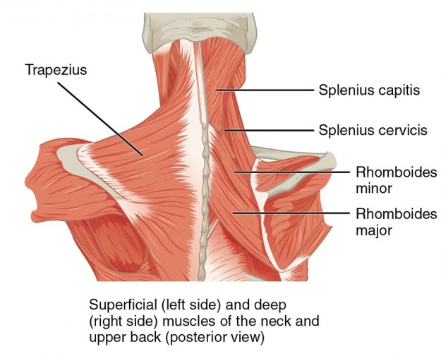 Topographic Anatomy Of The Back The Lecturio Medical Online Library
 Knee Joint Picture Image On Medicinenet Com
 Marc Joiret Webpage
 Figure 6 From The Menisco Femoral Ligaments Of The Human Knee Semantic Scholar
 Knee Human Anatomy Function Parts Conditions Treatments

 Knee Physiopedia
 Knee Leg Atlas Of Anatomy
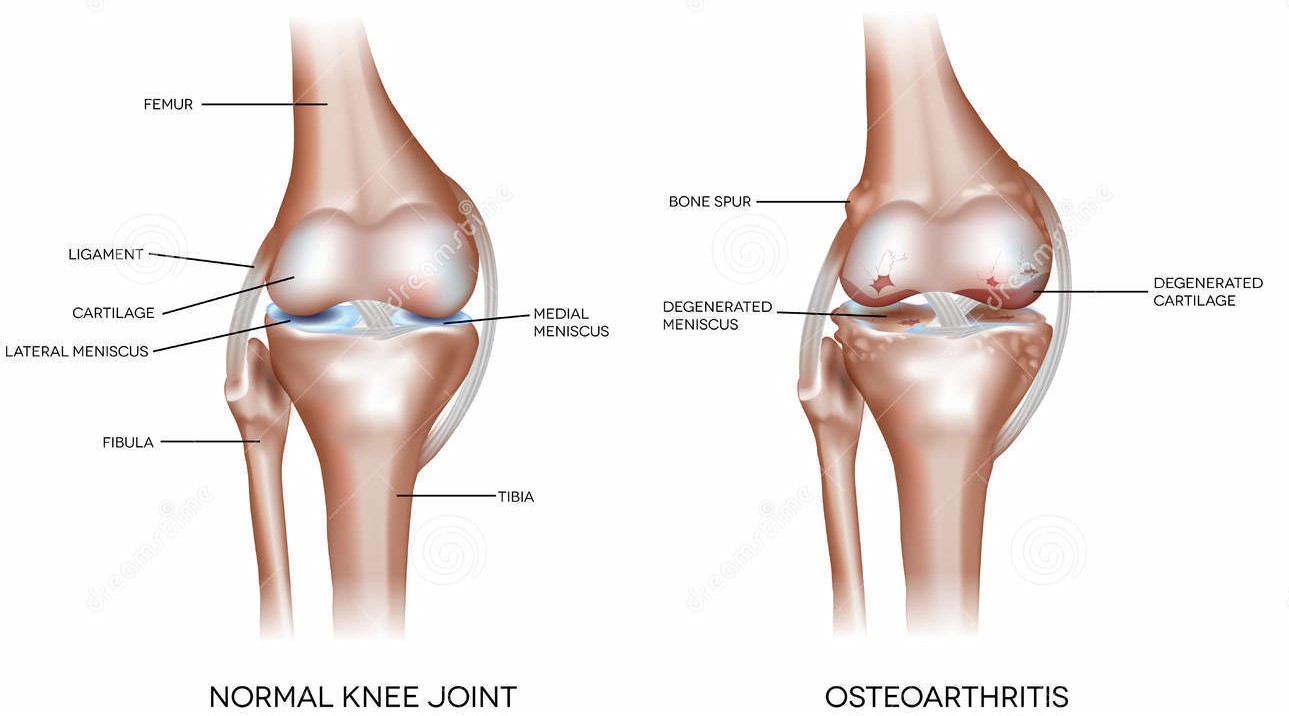
 Osteoarthritis And Normal Joint Stock Vector Illustration Of Care Ache 54350486
 Acl Dr Phil Huang
 Knee Joint Picture Image On Medicinenet Com
 Benefit Pt S Anatomy Series The Knee Part 2
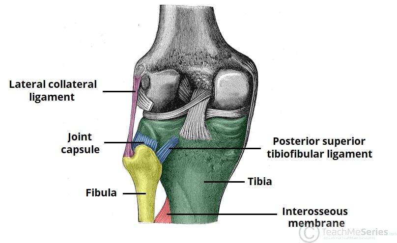 Tibiofibular Joints Proximal Distal Interosseous Membrane Teachmeanatomy
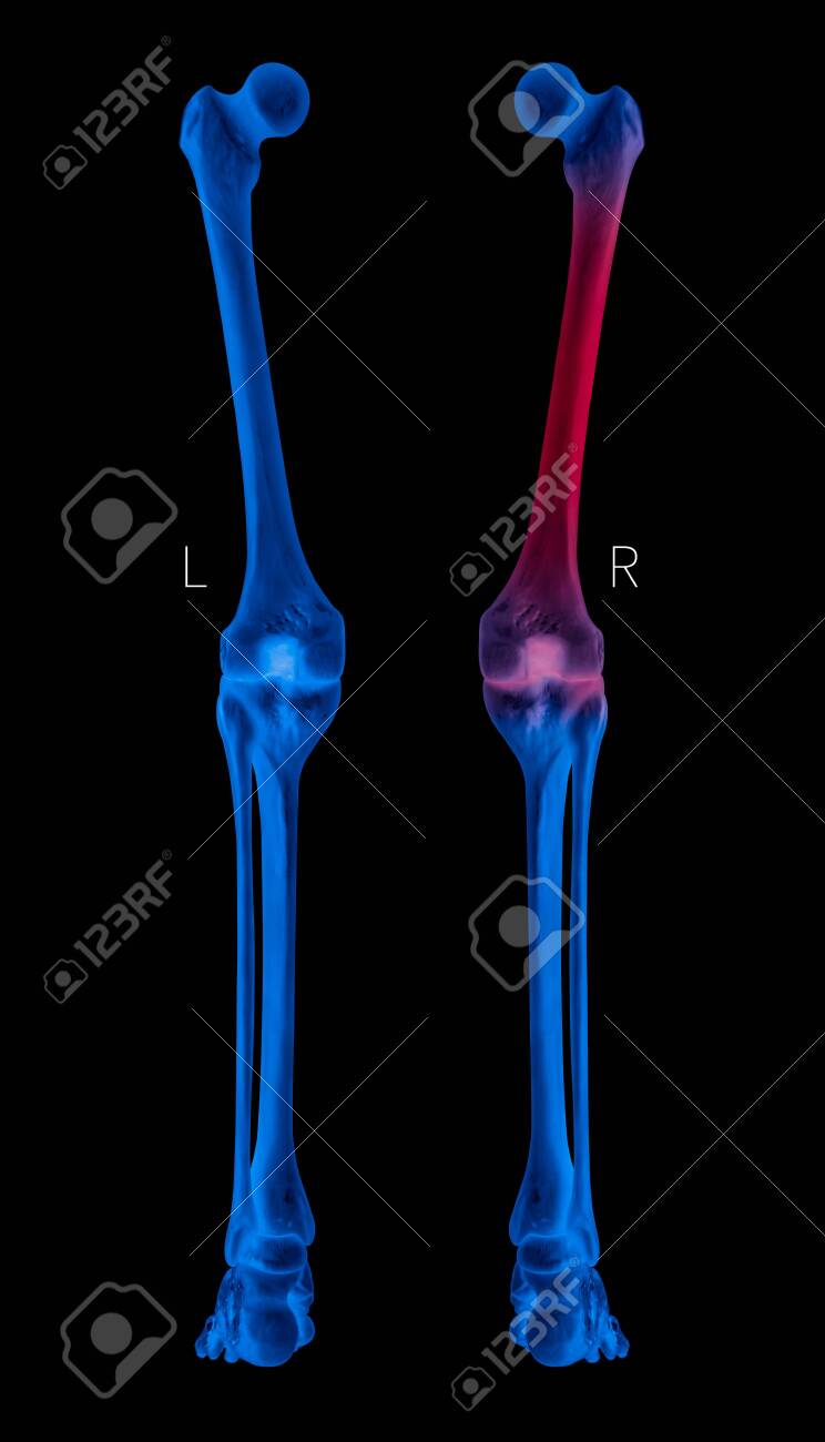 X Ray Of Human Leg Bone Left And Right Posterior View Red Highlights Stock Photo Picture And Royalty Free Image Image 121247653
 Knee Joint Anatomy Motion Knee Pain Explained
 Figure 3 From The Menisco Femoral Ligaments Of The Human Knee Semantic Scholar
![]() Knee Joint Anatomy Ligaments And Movements Kenhub
Knee Joint Anatomy Ligaments And Movements Kenhub
Https Encrypted Tbn0 Gstatic Com Images Q Tbn And9gctxxjaozuuss2y5xpz8b9bodf4efisgqumygjrye Kqrzylpykc Usqp Cau

 Anatomy Of Human Knee Joint Canvas Print By Stocktrekimages Redbubble
 Exam Series Guide To The Knee Exam Canadiem
 Popliteal Fossa Wikipedia
 Bones Advanced Anatomy 2nd Ed
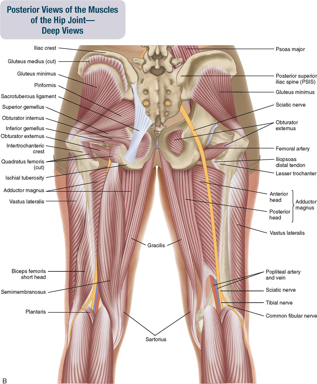 10 Muscles Of The Pelvis And Thigh Musculoskeletal Key
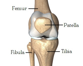
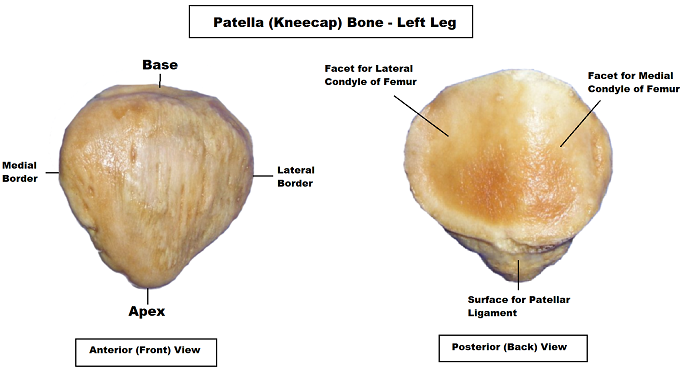 Patella Anatomy Patella Bone Anatomy Video And Notes
 Muscles Of The Pectoral Girdle And Upper Limbs Anatomy And Physiology I
 Anatomy Of The Elbow Joint Posterior Elbow View And Anterior Elbow View
 Anterior View And Posterior View Of The Human Leg Muscles Anatomy Www Anatomynote Com Calf Muscle Anatomy Body Muscle Anatomy Human Body Anatomy

Fractures Of The Proximal Tibia Shinbone Orthoinfo Aaos
 Anteromedial View Of Left Knee A The Superficial Medial Collateral Download Scientific Diagram
Https Drrobertlaprademd Com Wp Content Uploads 2015 07 Anatomy Of The Posterior Aspect Of The Knee 2007 Pdf
 Knee Anatomy Lateral View Human Anatomy

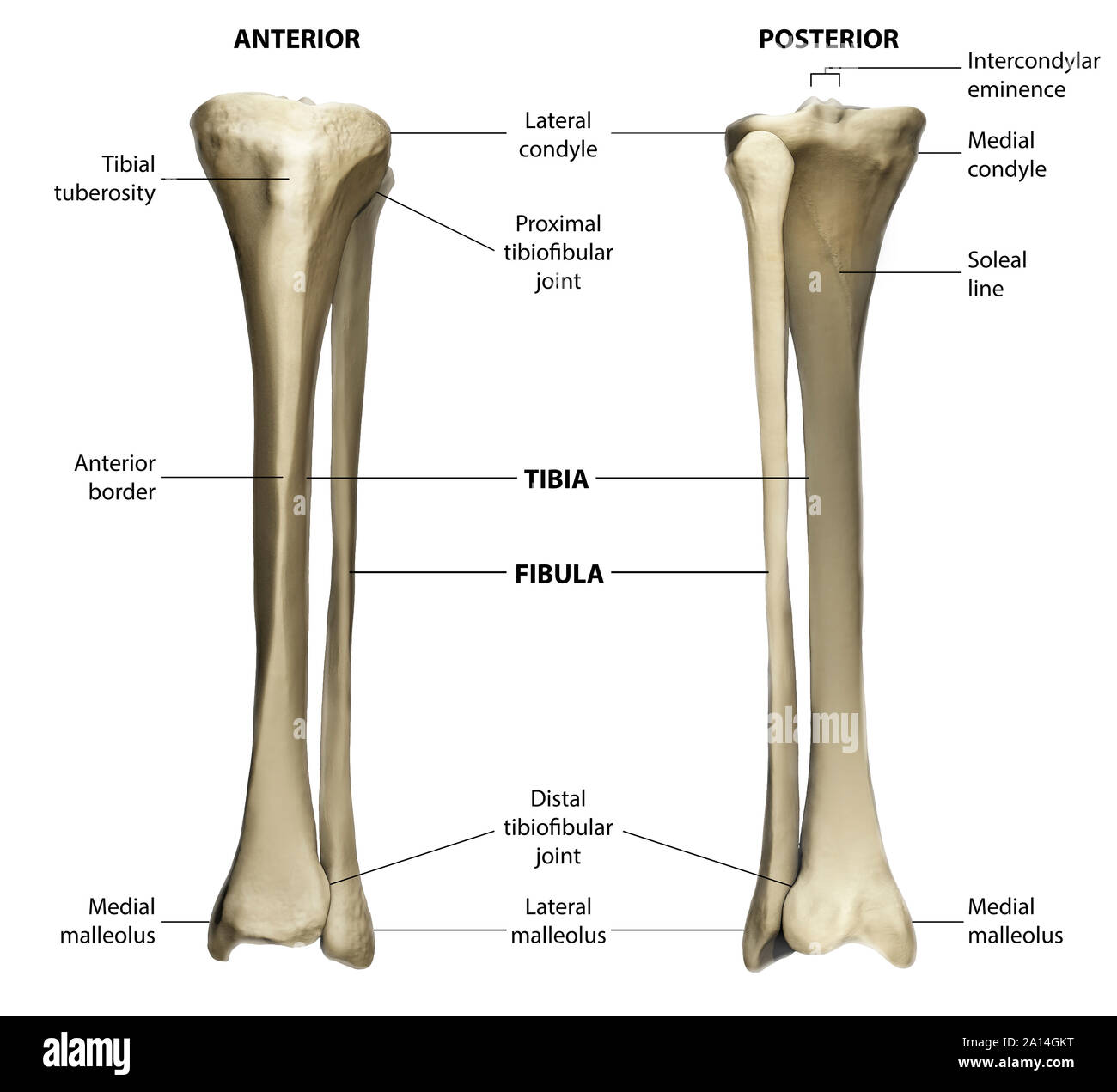 Anterior And Posterior View Of The Tibia And Fibula With Labeling Stock Photo Alamy
 Evaluation Of Patients Presenting With Knee Pain Part I History Physical Examination Radiographs And Laboratory Tests American Family Physician
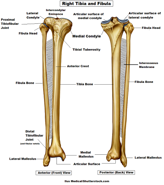 Tibia And Fibula Bone Anatomy
 Anatomical Chart Set Of Muscular Skeletal Anterior And Posterior Views 2 Matte Finish Charts Of Heavy Duty Paper Amazon Com Industrial Scientific
 Anatomy Acl Tear And Yoga Flying Elephant Yoga
Illustration Of Right Knee Posterior View Showing Bones Ligaments And Menisci Calisphere
 Right Knee Posterior And Sagittal Views Knee Posterior And Sagittal Views Knee Joint Posterior And Sagittal
 Muscles Of The Pectoral Girdle And Upper Limbs Anatomy And Physiology I
Https Encrypted Tbn0 Gstatic Com Images Q Tbn And9gcsxyw Ytujldnbkhmv5sldye9oaxsoxlqg6qzmbkuqqti0hnre0 Usqp Cau
 Knee Leg Atlas Of Anatomy
 Lateral Collateral Ligament Injury Of The Knee Physiopedia
 Semimembranosus Muscle An Overview Sciencedirect Topics
 Knee Pain